
level 8
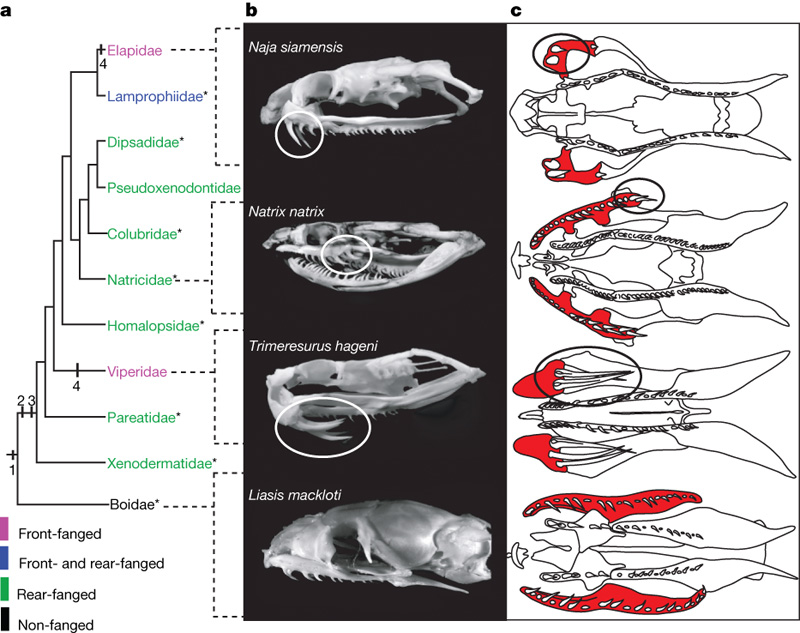 (a, Phylogeny from ref. 16. b, c, Adult skulls (Supplementary Table 4): lateral views (b); palate, schematic ventral views (c; maxilla colored, fangs circled). Asterisks indicate species studied by electron microscopy (Supplementary Fig. 5, Supplementary Table 3). The evolutionary changes leading from an unmodified maxillary dentition to the different fang types in advanced snakes are indicated at the nodes: (1) continuous maxillary dental lamina, no specialized sub regions—ancestral condition for advanced snakes; (2) evolution of posterior maxillary dental lamina—developmental uncoupling of posterior from anterior teeth; (3) starting differentiation of the posterior teeth with the venom gland; (4) loss of anterior dental lamina and development of front fangs. (Figure 1, Vonk 2008))
![[乖]](/static/emoticons/u4e56.png)
![[太开心]](/static/emoticons/u592au5f00u5fc3.png)
 (a–d, Palate, ventral view: top, anterior; scale bar, 0.5 mm; dotted lines, upper jaw (posterior margin of premaxilla to attachment of the mandible); boxes, schemes of maxillary odontogenic band (purple, shh expression; grey, no shh expression). Positions of fangs in b–d were identified histologically (Fig. 3, Supplementary Fig. 3). The odontogenic band in the front-fanged species is located posterior in the upper jaw (b, d). In the non-fanged outgroup (a) and the rear-fanged Natrix (c), the odontogenic band extends along the entire upper jaw. f, fang; mx, maxillary odontogenic band; pa, palatine odontogenic band; pt, pterygoid odontogenic band. e, Ontogenetic allometry in the fang in the front-fanged Causus displaces the fang along the upper jaw (Supplementary Figs 5–9, Supplementary Tables 5–9). Scale bars, 1 mm. We note the change in relative size of the upper jaw subregions: i, anterior; ii, fang; iii, posterior. d.a.o., days after oviposition. (Figure 2, Vonk 2oo8))
 (a–c, e–f, Sagittal sections, anterior to the left, of L. mackloti (Boidae) 22 d.a.o. (a), N. natrix (Natricidae) 22 d.a.o. (b), Calloselasma rhodostoma (Viperidae) 8 d.a.o. (c), N. natrix 22 d.a.o. (e), Naja siamensis (Elapidae) 23 d.a.o. (f). d, Transverse section, medial to the left, of Trimeresurus hageni (Viperidae) 8 d.a.o. The posterior maxillary dental laminae in b and e are similar in morphogenesis to the maxillary dental laminae in all front-fanged species examined (c, d, f; see also Fig. 4). Arrowheads, shh expression; amdl, anterior maxillary dental lamina; dr, dental ridge; e, eye; f, fang; mdl, maxillary dental lamina; pa, palatine dental lamina; pmdl, posterior maxillary dental lamina; t, tooth bud; vd, primordium of venom gland; scale bars, 300 microm. (Figure 3, Vonk 2008))
